Bipolar1Blog

Why nutritional psychiatry is the future of mental health treatment

Blueberries

Very interesting and may be so helpful.

Below is a direct quote from the article below. I wonder why we are not told to add vitamin and mineral as well as OTC anti-inflammatory supplementation to out diets and medicine regimen? It would be worth it to try it, if it doesn’t help, ok, but imagine it does help! I am going to ask my psychiatrist about all of these and if they have any adverse reactions with the medications I’m on. That’s would be the best way to go here. Wishing us all the best of physical and mental health.

“It is now known that many mental health conditions are caused by inflammation in the brain which ultimately causes our brain cells to die. This inflammatory response starts in our gut and is associated with a lack of nutrients from our food such as magnesium, omega-3 fatty acids, probiotics, vitamins and minerals that are all essential for the optimum functioning of our bodies. Recent research has shown that food supplements such as zinc, magnesium, omega 3, and vitamins B and D3 can help improve people’s mood, relieve anxiety and depression and improve the mental capacity of people with Alzheimer’s. Magnesium is one of most important minerals for optimal health, yet many people are lacking in it. One study found that a daily magnesium citrate supplement led to a significant improvement in depression and anxiety, regardless of age, gender or severity of depression. Improvement did not continue when the supplement was stopped. Omega-3 fatty acids are another nutrient that is critical for the development and function of the central nervous system – and a lack has been associated with low mood, cognitive decline and poor comprehension. Research has shown that supplements like zinc, magnesium and vitamins B and D can improve the mental capacity of people with Alzheimer’s. Shutterstock The role of probiotics – the beneficial live bacteria in your digestive system – in improving mental health has also been explored by psychiatrists and nutritionists, who found that taking them daily was associated with a significant reduction in depression and anxiety. Vitamin B complex and zinc are other supplements found to reduce the symptoms of anxiety and depression. Hope for the future? These over-the-counter” supplements are widely available in supermarkets, chemists and online health food stores, although the cost and quality may vary. For people who have not responded to prescription drugs or who cannot tolerate the side effects, nutritional intervention can offer hope for the future. There is currently much debate over the effectiveness of antidepressants. The use of food supplements offer an alternative approach that has the potential to make a significant difference to the mental health of all age groups.”


http://theconversation.com/why-nutritional-psychiatry-is-the-future-of-mental-health-treatment-92545

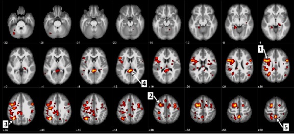
A lack of essential nutrients is known to contribute to the onset of poor mental health in people suffering from anxiety and depression, bipolar disorder, schizophrenia and ADHD. Nutritional psychiatry is a growing discipline that focuses on the use of food and supplements to provide these essential nutrients as part of an integrated or alternative treatment for mental health disorders. But nutritional approaches for these debilitating conditions are not widely accepted by mainstream medicine. Treatment options tend to be limited to official National Institute for Care Excellence (NICE) guidelines which recommend talking therapies and antidepressants. Use of antidepressants Antidepressant use has more than doubled in recent years. In England 64.7m prescriptions were issued for antidepressants in 2016 at a cost of £266.6m. This is an increase of 3.7m on the number of items prescribed in 2015 and more than double than the 31m issued in 2006. A recent Oxford University study found that antidepressants were more effective in treating depression than placebo. The study was led by Dr Andrea Cipriani who claimed that depression is under treated. Cipriani maintains that antidepressants are effective and a further 1m prescriptions should be issued to people in the UK. This approach suggests that poor mental health caused by social conditions is viewed as easily treated by simply dispensing drugs. But antidepressants are shunned by people whom they could help because of the social stigma associated with mental ill-health which leads to discrimination and exclusion. Prescriptions for 64.7m items of antidepressants were dispensed in England in 2016, the highest level recorded by the NHS. Shutterstock More worrying is the increase in the use of antidepressants by children and young people. In Scotland, 5,572 children under 18 were prescribed antidepressants for anxiety and depression in 2016. This figure has more than doubled since 2009/2010. But according to British psychopharmacologist Professor David Healy, 29 clinical trials of antidepressant use in young people found no benefits at all. These trials revealed that instead of relieving symptoms of anxiety and depression, antidepressants caused children and young people to feel suicidal. Healy also challenges their safety and effectiveness in adults. He believes that antidepressants are over-prescribed and that there is little evidence that they are safe for long-term use. Antidepressants are said to create dependency, have unpleasant side effects and cannot be relied upon to always relieve symptoms. Nutrition and poor mental health In developed countries such as the UK we eat a greater variety of foodstuffs than ever before – but it doesn’t follow that we are well nourished. In fact, many people do not eat enough nutrients that are essential for good brain health, opting for a diet of heavily processed food containing artificial additives and sugar. The link between poor mental health and nutritional deficiencies has long been recognised by nutritionists working in the complementary health sector. However, psychiatrists are only now becoming increasingly aware of the benefits of using nutritional approaches to mental health, calling for their peers to support and research this new field of treatment. It is now known that many mental health conditions are caused by inflammation in the brain which ultimately causes our brain cells to die. This inflammatory response starts in our gut and is associated with a lack of nutrients from our food such as magnesium, omega-3 fatty acids, probiotics, vitamins and minerals that are all essential for the optimum functioning of our bodies. Recent research has shown that food supplements such as zinc, magnesium, omega 3, and vitamins B and D3 can help improve people’s mood, relieve anxiety and depression and improve the mental capacity of people with Alzheimer’s. Magnesium is one of most important minerals for optimal health, yet many people are lacking in it. One study found that a daily magnesium citrate supplement led to a significant improvement in depression and anxiety, regardless of age, gender or severity of depression. Improvement did not continue when the supplement was stopped. Omega-3 fatty acids are another nutrient that is critical for the development and function of the central nervous system – and a lack has been associated with low mood, cognitive decline and poor comprehension. Research has shown that supplements like zinc, magnesium and vitamins B and D can improve the mental capacity of people with Alzheimer’s. Shutterstock The role of probiotics – the beneficial live bacteria in your digestive system – in improving mental health has also been explored by psychiatrists and nutritionists, who found that taking them daily was associated with a significant reduction in depression and anxiety. Vitamin B complex and zinc are other supplements found to reduce the symptoms of anxiety and depression. Hope for the future? These over-the-counter” supplements are widely available in supermarkets, chemists and online health food stores, although the cost and quality may vary. For people who have not responded to prescription drugs or who cannot tolerate the side effects, nutritional intervention can offer hope for the future. There is currently much debate over the effectiveness of antidepressants. The use of food supplements offer an alternative approach that has the potential to make a significant difference to the mental health of all age groups. The emerging scientific evidence suggests that there should be a bigger role for nutritional psychiatry in mental health within conventional health services. If the burden of mental ill health is to be reduced, GPs and psychiatrists need to be aware of the connection between food, inflammation and mental illness. Medical education has traditionally excluded nutritional knowledge and its association with disease. This has led to a situation where very few doctors in the UK have a proper understanding of the importance of nutrition. Nutritional interventions are thought to have little evidence to support their use to prevent or maintain well-being and so are left to dietitians, rather than doctors, to advise on. But as the evidence mounts up, it is time for medical education to take nutrition seriously so that GPs and psychiatrists of the future know as much about its role in good health as they do about anatomy and physiology. The state of our mental health could depend on it.

IBM’s AI can predict schizophrenia by looking at the brain’s blood flow

With 74% accuracy!

https://www.engadget.com/2017/07/20/ibms-ai-can-predict-schizophrenia-by-looking-at-the-brains-blo/?sr_source=Twitter

IBM’s AI can predict schizophrenia by looking at the brain’s blood flow

And it does so with 74 percent accuracy

Schizophrenia is not a particularly common mental health disorder in America, affecting just 1.2 percent of the population (around 3.2 million people), but its effects can be debilitating. However, pioneering research conducted by IBM and the University of Alberta could soon help doctors diagnose the onset of the disease and the severity of its symptoms using a simple MRI scan and a neural network built to look at blood flow within the brain.

“This unique, innovative multidisciplinary approach opens new insights and advances our understanding of the neurobiology of schizophrenia, which may help to improve the treatment and management of the disease,” Dr. Serdar Dursun, a Professor of Psychiatry & Neuroscience with the University of Alberta, said in a statement.

MRI scans showing statistically significant differing blood flows within the brain – Image: IBM

The research team first trained its neural network on a 95-member dataset of anonymized fMRI images from the Function Biomedical Informatics Research Network which included scans of both patients with schizophrenia and a healthy control group. These images illustrated the flow of blood through various parts of the brain as the patients completed a simple audio-based exercise. From this data, the neural network cobbled together a predictive model of the likelihood that a patient suffered from schizophrenia based on the blood flow. It was able to accurately discern between the control group and those with schizophrenia 74 percent of the time.

“We’ve discovered a number of significant abnormal connections in the brain that can be explored in future studies,” Dursun continued, “and AI-created models bring us one step closer to finding objective neuroimaging-based patterns that are diagnostic and prognostic markers of schizophrenia.”

What’s more, the model managed to also predict the severity of symptoms once they set in. These insights could lead researchers to more effective diagnostic tools and treatment options. And why wouldn’t they? IBM’s most famous AI, Watson, has already shown that neural networks are surprisingly adept at coming up with effective cancer treatment regimens.


Immune Activity During Pregnancy Tied to Neuronal Defects, Anxiety, and Cognitive Impairments

Apple blossoms

I’m definitely cataloguing this in the Interesting category!

Neurons called Parvalbumin Interneurons in the brains of mice pups were underactive when their mothers had an infection while pregnant, i.e. the mother’s immune systems were ramped up due to the infection. This did not happen to mice whose mothers had no infection during pregnancy. The mice with underactive Parvalbumin Interneurons exhibited more anxiety and struggled with behavioral tests.

Also it was possible to induce these effects (anxiety and difficulty in behavioral tests) in normal mice (no infection in mother while pregnant) by shutting off the Parvalbumin Interneurons.

Parvalbumin Interneurons are neurons which send inhibitory signals to other neurons are much weaker in neurons of mice born to mothers whose immune system had been activated during pregnancy. “Parvalbumin interneurons help coordinate the activity of other cells in the brain, and are thought to be important for memory and cognitive flexibility. Reduced numbers and structural abnormalities in parvalbumin interneurons have been linked to multiple psychiatric disorders…”

https://bbrfoundation.org/brain-matters-discoveries/immune-activity-during-pregnancy-tied-to-neuronal-defects-anxiety-and

Researchers are investigating the possibility that infections during pregnancy increase the likelihood that the fetus will develop into a person who will have mental illness later in life. There is evidence that immune activity in response to maternal infections may increase the offspring’s risk of schizophrenia,bipolar disorder, depression, and anxiety disorders.

In a new animal study led by Christoph Kellendonk, Ph.D., a 2002 and 2008 NARSAD Young Investigator at Columbia University Medical Center, scientists have shown that inhibitory brain cells called parvalbumin interneurons are particularly vulnerable to such maternal immune activation. These cells do not signal as they should in mice whose mothers’ immune systems are activated during pregnancy, the researchers have shown. What’s more, the signaling problems are associated with cognitive impairments and anxiety-like behavior in mice.

Activated immune system in pregnant mice linked to later cognitive impairments in their offspring. Tweet >

The research team, which included Alan Stewart Brown, M.D., M.P.H., a 1993, 1996 Young Investigator, 2000, 2004 Independent Investigator and 2015 Distinguished Investigator, 2013 Young Investigator Sarah E. Canetta, Ph.D. and BBRF Scientific Council Member and 2001, 2003 Young Investigator Joshua A. Gordon, M.D., Ph.D., all at Columbia, published its findings February 2 in the journal Molecular Psychiatry.

Parvalbumin interneurons help coordinate the activity of other cells in the brain, and are thought to be important for memory and cognitive flexibility. Reduced numbers and structural abnormalities in parvalbumin interneurons have been linked to multiple psychiatric disorders, but so far it has been difficult to assess how these abnormalities affect brain function.

In their mouse study, Dr. Kellendonk and colleagues determined that the inhibitory signals that parvalbumin interneurons usually send to target cells are much weaker than usual in mice whose mothers’ immune systems had been active during pregnancy. Those mice struggled with a behavioral test that involved task switching, suggesting certain cognitive impairments, and also exhibited more anxiety than mice whose mothers had no immune activation during pregnancy.

In mice whose mothers did not have activated immune systems during pregnancy, the scientists could provoke the same increase in anxiety and cognitive defects simply by artificially shutting off parvalbumin interneurons, supporting the idea that defects in the cells were responsible for the affected animals’ behavior.

Takeaway: Study in mice identifies brain cells that are vulnerable to a mother’s infection during pregnancy. Reduced signaling from these cells was associated with increased anxiety and cognitive impairments.

It’s the Immune System!

DSCN5325

In normal brains, the number of synapses (connections between neurons where neurotransmitters act and brain activity takes place and nerve impulses and information is passes on) is pruned or  whittled down as the brain matures from the womb to adolescence. These synapses are eliminated by immune cells of the brain called microglia.

First the hypothesis that Schizophrenia is caused by activation of microglia which eat away at synapses in childhood or adolescence, leading to fewer synapses and this mental illness! An amazing paper about which I wrote a post (https://wordpress.com/post/bipolar1blog.wordpress.com/3623) C1q is a protein that tags the neuronal synapses, once a synapse is tagged, microglia (the immune cells of the brain) come and chomp it away, voila, no more synapse. The information that that one synapse was transmitting is now lost. If this happens to many, many, synapses, a lot of communication and information is lost. And this loss leads to schizophrenia!

Now the same observations about Alzheimer’s as well! Microglia are eating away too may synapses in areas of the brain that are key to memory. β Amyloid is a plaque of protein found to a much larger extent in the brains of people with Alzheimer’s. It is a deposit that is seen along neurons of people with Alzheimer’s. What this research team has found is that C1q in conjunction with the existence of β Amyloid plaques is what causes the microglia to eat up healthy synapses. This lead to destruction of brain cell connectivity, especially in the areas that house memory. So this process that happens naturally in the womb, somehow gets turned on later in life and causes pruning of connections in neurons which we need and leads to Alzheimer’s.

Again, it’s the immune system stupid!

http://www.sciencemag.org/news/2016/03/over-pruning-synapses-may-drive-early-stage-alzheimer-s-disease?utm_source=newsfromscience&utm_medium=facebook-text&utm_campaign=alzhapses-3346

Alzheimer’s may be caused by haywire immune system eating brain connections

More than 99% of clinical trials for Alzheimer’s drugs have failed, leading many to wonder whether pharmaceutical companies have gone after the wrong targets. Now, research in mice points to a potential new target: a developmental process gone awry, which causes some immune cells to feast on the connections between neurons.

“It is beautiful new work,” which “brings into light what’s happening in the early stage of the disease,” says Jonathan Kipnis, a neuroscientist at the University of Virginia School of Medicine in Charlottesville.

Most new Alzheimer’s drugs aim to eliminate β amyloid, a protein that forms telltale sticky plaques around neurons in people with the disease. Those with Alzheimer’s tend to have more of these deposits in their brains than do healthy people, yet more plaques don’t always mean more severe symptoms such as memory loss or poor attention, says Beth Stevens of Boston Children’s Hospital, who led the new work.

What does track well with the cognitive decline seen in Alzheimer’s disease—at least in mice that carry genes that confer high risk for the condition in people—is a marked loss of synapses, particularly in brain regions key to memory, Stevens says. These junctions between nerve cells are where neurotransmitters are released to spark the brain’s electrical activity.

Stevens has spent much of her career studying a normal immune mechanism that prunes weak or unnecessary synapses as the brain matures from the womb through adolescence, allowing more important connections to become stronger. In this process, a protein called C1q sets off a series of chemical reactions that ultimately mark a synapse for destruction. After a synapse has been “tagged,” immune cells called microglia—the brain’s trash disposal service—know to “eat” it, Stevens says. When this system goes awry during the brain’s development, whether in the womb or later during childhood and into the teen years, it may lead to psychiatric disorders such as schizophrenia, she says.

Stevens hypothesized that the same mechanism goes awry in early Alzheimer’s disease, leading to the destruction of good synapses and ultimately to cognitive impairment. Using two Alzheimer’s mouse models—each of which produces excess amounts of the β amyloid protein, and develops memory and learning impairments as they age—she and her team found that both strains had elevated levels of C1q in brain tissue. When they used an antibody to block C1q from setting off the microglial feast, however, synapse loss did not occur, the team reports today in Science.

To Stevens, that suggests that the normal mechanism for pruning synapses during development somehow gets turned back on again in the adult brain in Alzheimer’s, with dangerous consequences. “Instead of nicely whittling away [at synapses], microglia are eating when they’re not supposed to,” she says.

The group is now tracking these mice to see whether a drug that blocks C1q slows their cognitive decline. To determine whether elevated β amyloid can cause the C1q system to go haywire, Stevens and colleagues also injected a form of the protein which is known to generate plaques into the brains of normal mice and so-called knockouts that could not produce C1q because of a genetic mutation. Although normal mice exposed to the protein lost many synapses, knockouts were largely unaffected, Stevens says. In addition, microglia only went after synapses when β amyloid was present, suggesting that the combination of protein and C1q is what destroys synapses, rather than either element alone, she says, adding that other triggers, such as inflammatory molecules called cytokines, might also set the system off.

The findings contradict earlier theories which held that increased microglia and C1q activity were merely part of an inflammatory reaction to β amyloid plaques. Instead, microglia seem to start gorging on synapses long before plaques form, Stevens says. She and several co-authors are shareholders in Annexon Biosciences, a biotechnology company that will soon start testing the safety of a human form of the antibody the team used to block C1q, known as ANX-005, in people.

Such a central role for microglia in Alzheimer’s disease is “still on the controversial side,” says Edward Ruthazer, a neuroscientist at the Montreal Neurological Institute and Hospital in Canada. One “really compelling” sign that the mechanism is important in people would be if high levels of C1q in cerebrospinal fluid early on predicted developing full-blown Alzheimer’s later in life, he says. Still, he says, “it’s difficult to argue with the strength of the study’s evidence.”

Scientists Move Closer to Understanding Schizophrenia’s Cause (Amazing research and information!)

SCHIZOPHRENIA
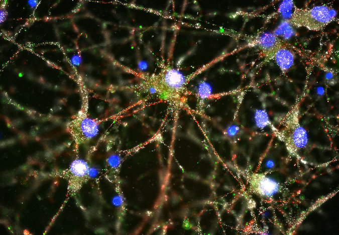
This is absolutely amazing! Scientists have found out that the cause of schizophrenia is too much synaptic pruning of neurons, “in which the brain sheds weak or redundant connections between neurons as it matures” in the prefrontal cortex. This begins in adolescence and the symptoms of schizophrenia also start in adolescence! “People who carry genes that accelerate or intensify that pruning are at higher risk of developing schizophrenia than those who do not, the new study suggests.”

They knew that the MHC locus was involved in schizophrenia, they pinpointed the exact gene that is involved, the C-4 gene, this is the gene that facilitates the aggressive tagging of connections, thereby accelerating pruning. Wow, this is so amazing! Here it is in a nutshell, why people get schizophrenia! I am totally floored! So floored, I almost forgot to put the reference, but here is is:

http://www.nytimes.com/2016/01/28/health/schizophrenia-cause-synaptic-pruning-brain-psychiatry.html

“Scientists reported on Wednesday that they had taken a significant step toward understanding the cause of schizophrenia, in a landmark study that provides the first rigorously tested insight into the biology behind any common psychiatric disorder.

More than two million Americans have a diagnosis of schizophrenia, which is characterized by delusional thinking and hallucinations. The drugs available to treat it blunt some of its symptoms but do not touch the underlying cause.

The finding, published in the journal Nature, will not lead to new treatments soon, experts said, nor to widely available testing for individual risk. But the results provide researchers with their first biological handle on an ancient disorder whose cause has confounded modern science for generations. The finding also helps explain some other mysteries, including why the disorder often begins in adolescence or young adulthood.

“They did a phenomenal job,” said David B. Goldstein, a professor of genetics at Columbia University who has been critical of previous large-scale projects focused on the genetics of psychiatric disorders. “This paper gives us a foothold, something we can work on, and that’s what we’ve been looking for now, for a long, long time.”

The researchers pieced together the steps by which genes can increase a person’s risk of developing schizophrenia. That risk, they found, is tied to a natural process called synaptic pruning, in which the brain sheds weak or redundant connections between neurons as it matures. During adolescence and early adulthood, this activity takes place primarily in the section of the brain where thinking and planning skills are centered, known as the prefrontal cortex. People who carry genes that accelerate or intensify that pruning are at higher risk of developing schizophrenia than those who do not, the new study suggests.

Some researchers had suspected that the pruning must somehow go awry in people with schizophrenia, because previous studies showed that their prefrontal areas tended to have a diminished number neural connections, compared with those of unaffected people. The new paper not only strongly supports that this is the case, but also describes how the pruning probably goes wrong and why, and identifies the genes responsible: People with schizophrenia have a gene variant that apparently facilitates aggressive “tagging” of connections for pruning, in effect accelerating the process.

Some scientists warned that the history of biological psychiatry stands as a caution against premature optimism. “This work is extremely persuasive,” said Dr. Samuel Barondes, a professor of psychiatry at the University of California, San Francisco, “but any step forward is not only rare and unusual, it’s just one step in a journey of a thousand miles” to improved treatments.

The study, by scientists from Harvard Medical School, Boston Children’s Hospital and the Broad Institute, a research center allied with Harvard and the Massachusetts Institute of Technology, provides a showcase of biomedical investigation at its highest level. The research team began by focusing on a location on the human genome, the MHC, which was most strongly associated with schizophrenia in previous genetic studies. On a bar graph — called a Manhattan plot because it looks like a cluster of skyscrapers — the MHC looms highest.


“The MHC is the Freedom Tower” of the Manhattan plot, said Eric S. Lander, the director of the Broad Institute. “The question was, what’s in there?”

The area is a notoriously dark warren in the genome known to contain genes that facilitate the body’s immune response, for example, by flagging invading bacteria to be destroyed. That property had given rise to speculation that schizophrenia might be a kind of autoimmune condition, in which the body attacked its own cells.

But the research team, led by Steven McCarroll, an associate professor of genetics at Harvard, and by Aswin Sekar, one of his graduate students, found something different. Using advanced statistical methods, the team found that the MHC locus contained four common variants of a gene called C4, and that those variants produced two kinds of proteins, C4-A and C4-B.

The team analyzed the genomes of more than 64,000 people and found that people with schizophrenia were more likely to have the over-active forms of C4-A than control subjects. “C4-A seemed to be the gene driving risk for schizophrenia,” Dr. McCarroll said, “but we had to be sure.”

The researchers turned to Beth Stevens, an assistant professor of neurology at Boston Children’s Hospital and Harvard, who in 2007 was an author of a study showing that the products of MHC genes were involved in synaptic pruning in normal developing brains. But how important was this C4 protein, exactly? Very important, it turned out: Mice bred without the genes that produce C4 showed clear signs that their synaptic pruning had gone awry, Dr. Stevens’s lab showed.

Taken together, Dr. Stevens said in an interview, “the evidence strongly suggested that too much C4-A leads to inappropriate pruning during this critical phase of development.”

In particular, the authors concluded, too much C4-A could mean too much pruning — which would explain not only the thinner prefrontal layers in schizophrenia, but also the reason that the disorder most often shows itself in people’s teenage years or early twenties. “The finding connects all these dots, all these disconnected observations about schizophrenia, and makes them make sense,” Dr. McCarroll said.

Carrying a gene variant that facilitates aggressive pruning is hardly enough to cause schizophrenia; far too many other factors are at work. Having such a variant, Dr. McCarroll estimates, would increase a person’s risk by about 25 percent over the 1 percent base rate of schizophrenia — that is, to 1.25 percent. That is not nearly enough to justify testing in the general population, even if further research confirms the new findings and clarifies the roles of other associated genes.

Yet the equation changes when it comes to young people who are at very high risk of developing the disorder, because they are showing early signs — a sudden slippage in mental acuity and memory, or even internal “voices” that seem oddly real. This ominous period may last a year or more, and often does not lead to to full-blown schizophrenia. The researchers hope that the at-risk genetic profile, once it has been fleshed out more completely, will lead to the discovery of biomarkers that could help clarify a prognosis in these people.

Developing a drug to slow or modulate pruning poses another kind of challenge. If the new study shows anything, it is that synaptic pruning is a delicate, exquisitely timed process, and that it is still poorly understood. The team does not yet know, for example, why C4-A leads to a different rate or kind of pruning than C4-B. Any medication that tampered with that system would be a risky proposition, the authors and outside experts agreed.

“We’re all very excited and proud of this work,” Dr. Lander said. “But I’m not ready to call it a victory until we have something that can help patients.”

A genome-wide association study of kynurenic acid in cerebrospinal fluid: implications for psychosis and cognitive impairment in bipolar disorder


A variant of the SNX7 gene and its reduced expression, is associated with levels of a protein called kynurenic acid aka KYNA (an end metabolite in tryptophan metabolism.) This is associated with the psychotic (out of touch with reality) symptoms and cognitive impairment seen in bipolar disorder and schizophrenia. Interestingly, this pathway involves signaling via the immune cells (glia) of the brain! Another immune cell connection to mental illness! And KYNA could be targeted for drug development, as reducing it should lead to a decrease in psychotic symptoms as well as cognitive impairment.

http://www.nature.com/mp/journal/vaop/ncurrent/full/mp2015186a.html

Abstract

Elevated cerebrospinal fluid (CSF) levels of the glia-derived N-methyl-d-aspartic acid receptor antagonist kynurenic acid (KYNA) have consistently been implicated in schizophrenia and bipolar disorder. Here, we conducted a genome-wide association study based on CSF KYNA in bipolar disorder and found support for an association with a common variant within 1p21.3. After replication in an independent cohort, we linked this genetic variant—associated with reduced SNX7 expression—to positive psychotic symptoms and executive function deficits in bipolar disorder. A series of post-mortem brain tissue and in vitro experiments suggested SNX7 downregulation to result in a caspase-8-driven activation of interleukin-1β and a subsequent induction of the brain kynurenine pathway. The current study demonstrates the potential of using biomarkers in genetic studies of psychiatric disorders, and may help to identify novel drug targets in bipolar disorder.

Top

Introduction

Elevation of brain kynurenic acid (KYNA) is a consistently found biochemical aberration in schizophrenia and bipolar disorder (BD).1, 2, 3, 4, 5, 6, 7 Brain KYNA is mainly produced in astrocytes as an end-metabolite of the kynurenine pathway of tryptophan metabolism. This pathway is highly inducible by inflammatory stimuli,8 and we have previously reported that cerebrospinal fluid (CSF) levels of the proinflammatory cytokine interleukin (IL)-1β are markedly increased in patients with BD or schizophrenia, although the majority of other cytokines measured in this study were undetectable.9, 10

KYNA is a neuroactive metabolite that antagonizes the glycine co-agonist site of the N-methyl-d-aspartic acid receptor (NMDAR).8 Administration of synthetic NMDAR antagonists causes psychotic symptoms in healthy individuals,11 and exacerbates psychotic features in patients with schizophrenia.12 Psychotic symptoms are core features of schizophrenia, and more than half of patients with BD will experience psychosis in their lifetime.13 Supporting that KYNA might be specifically involved in the pathophysiology underlying psychotic symptoms, we have found higher levels of CSF KYNA in BD-I patients with a history of psychosis compared with those who had never experienced psychosis.14 KYNA also noncompetitively antagonizes the cholinergic α7 nicotinic receptor, and animal studies indicate that increased brain KYNA might cause cognitive deficits.8 In rats, increased brain KYNA causes behavioral responses analogous to impaired set-shifting in humans,15 an index of executive function. Set-shifting dysfunction as measured by the trail making test (TMT) is indeed a feature of schizophrenia and euthymic BD,16, 17 especially in BD patients with a history of psychosis.18

Family history is the strongest risk factor for BD, but an important obstacle for progress in psychiatric genetics is that psychiatric syndromes—based solely on symptom clustering—do not necessarily reflect specific underlying biological dysfunctions and may be insufficient to delineate heritable phenotypes.19 Indeed, epidemiological and molecular genetic studies have blurred the diagnostic boundary between schizophrenia and BD by demonstrating that these disorders have partly shared genetic causes.20, 21 Complementary approaches to unearth causal genetic mutations are therefore needed. One approach is to focus on biomarkers, that is, measurable key components in biological pathways between genotype and disease.22 For this purpose, the use of CSF KYNA may be particularly rewarding given its biological links to distinct subdomains of pathology present in both BD and schizophrenia.

In this study of euthymic BD patients, we found CSF IL-1β and KYNA to be associated with a history of psychosis and set-shifting impairment. CSF levels of KYNA were also strongly associated with the dopamine metabolite homovanillic acid (HVA). We conducted a genome-wide association study (GWAS) against CSF levels of KYNA in BD that revealed a genome-wide significant association with the single-nucleotide polymorphism (SNP) rs10158645 within 1p21.3, a finding that was replicated in an independent cohort of BD patients. Furthermore, we analyzed this SNP in relation to CSF HVA, a history of psychosis (followed by a replication in a large data set of 565 BD patients) and set-shifting ability. As the minor allele in rs10158645 was associated with decreased expression of sorting nexin 7 (SNX7), we attempted to decipher the biochemical chain of events using a multipronged approach including causal inference analyses of clinical data, post-mortem data and cell culture studies. These experiments converged on the proposal that decreased SNX7 expression is linked to increased CSF KYNA concentration and ultimately psychosis and set-shifting difficulties in BD through caspase-8-driven activation of IL-1β.


Negative Symptoms

SCHIZO

This is a graphic for negative (meaning things that are absent or missing, such as affect, pleasure, speaking, and activity) symptoms of schizophrenia, but these very symptoms are also present in the depressive phase of bipolar disorder. And these can also be erroneously attributed to laziness, “just not trying enough”, or unwillingness, but they are due to illness.

Schizophrenia, bipolar disorder and major depression share genetic risk factors: Study

DSCN6809

From the article below. Again, immune involvement. “The researchers found strong associations between mechanisms related to immune function and changes in processes when genes are turned on and off. The findings confirm known mechanisms as well as revealing new ones that pertain to the development of psychiatric disorders.”

I know when I am in full blown mania and out of touch with reality, there is no difference between me and a person who has schizophrenia. The thing is that my getting to that point can be prevented by taking Lithium (for me), whereas a person with schizophrenia has a lot more trouble coming out from delusions, hallucinations and back in touch with reality.

http://www.belmarrahealth.com/schizophrenia-bipolar-disorder-and-major-depression-share-genetic-risk-factors-study/

Schizophrenia, bipolar disorder and major depression have been found to share a genetic risk factor, according to a new study. Aside from the recent research, many previous studies also showed a genetic link between all three mental disorders. Below are synopses of the health studies that reveal the connection they all possess.

Study on shared genetic risk factors for schizophrenia, bipolar disorder and major depression

Research published in Nature Neuroscience from the Louisiana State University Health Science Centers revealed a genetic risk factor that is shared between schizophrenia, bipolar disorder and major depression. Lead researcher, Nancy Buccola, and her team examined data from 60,000 participants, including those with schizophrenia, bipolar disorder, major depression, autism, attention deficit disorders as well as individuals without any diagnosed conditions.

Study on shared genetic risk factors for schizophrenia, bipolar disorder and major depressionThe researchers found strong associations between mechanisms related to immune function and changes in processes when genes are turned on and off. The findings confirm known mechanisms as well as revealing new ones that pertain to the development of psychiatric disorders.

Treatments are available for many mental disorders but many patients do not obtain relief from such treatments. Buccola stated, “The PGC is a collaboration of some of the finest psychiatric genetic researchers in the world who are working together to understand the biology that underlies psychiatric disorders. This knowledge is critical in developing more effective and personalized treatments. I feel fortunate to make even a small contribution to this important work.”

Previous study shows schizophrenia and bipolar disorder cause dendritic spine loss in brain

Alternative research has found that schizophrenia and bipolar disorder both play a role in dendritic spine loss in the brain. The findings suggest that the two disorders share similar pathopsychological features.

Dendritic spines play a role in many brain functions. To achieve their results, researchers looked at individuals with schizophrenia, bipolar disorder, and individuals not affected by either disorder.

Spine density was reduced in those with bipolar disorder and those with schizophrenia, when compared to the control group. Furthermore, there was a significant reduction in spines per dendrite in both bipolar individuals and schizophrenics.

Lead researcher, Glenn T. Konopaske, M.D., said, “The current study suggests that spine pathology is common to both [schizophrenia] and [bipolar]. Moreover, the study of the mechanisms underlying the spine pathology might reveal additional similarities and differences between the two disorders, which could lead to the development of novel biomarkers and therapeutics.”

Bipolar disorder is often misdiagnosed as major depressive disorder (MDD)

Bipolar disorder is often misdiagnosed as major depressive disorder (MDD)Research has shown that bipolar disorder is often misdiagnosed as major depressive disorder (MDD). In bipolar disorder, individuals experience intense lows in mood and euphoric highs. In major depressive disorder, individuals experience steady, intense lows in mood. Because episodes of low mood can last for days or even weeks in those with bipolar, it can lead to a misdiagnosis of major depressive disorder. Researchers are closing in on an objective to help distinguish between the two conditions in order to reduce misdiagnosis.

Current diagnostic methods involve interviews with the patient, but this can be subjective and misleading. Researchers decided to combine techniques together in order to create a more accurate diagnostic method. The techniques used are gas chromatography-mass spectrometry and nuclear magnetic resonance, which analyze the urine of patients with MDD and bipolar disorder in order to uncover biomarkers of each disorder. These biomarkers will allow doctors to improve diagnosis by 89 to 91 percent.

Depression in patients with schizophrenia

One study found that a quarter (25 percent) of those with schizophrenia also have course-related depression. Depression in schizophrenia patients is related to a reduction in social and vocational functioning and also increases the risk of a psychotic relapse.

Depression in schizophrenia often has poor outcomes; patients have more suicidal thoughts, suicide attempts, and suicides.

It can be difficult to diagnose depression and schizophrenia separately as the “negative” symptoms related to schizophrenia can present themselves like depression. Negative symptoms refer to social withdrawal, low motivation and energy, difficulty experiencing pleasure or having interests and an impaired thought process; all symptoms seen in depression as well.

For the many similarities presented in both schizophrenia and depression, not only is distinguishing between the two difficult, but depression can often be seen in many schizophrenic patients as well.

From all the presented studies we see many links and associations between schizophrenia, bipolar disorder and depression. By continuing to make these links, we can obtain a better understanding of these mental disorders, which could greatly assist in developing more specific treatments that could offer more patients greater relief.

Brain fold tied to hallucinations

http://mobile.the-scientist.com/article/44547/brain-fold-tied-to-hallucinationsThe Scientist

The NutshellThe Scientist

Brain Fold Tied to Hallucinations

A shorter crease in the medial prefrontal cortex is linked with a higher risk of schizophrenics experiencing hallucinations.

By Kerry Grens | November 19, 2015
WIKIMEDIA, DATABASE CENTER FOR LIFE SCIENCE

People with schizophrenia who experience hallucinations are more likely to have a certain contour to their brain—specifically, a shorter groove in the medial prefrontal cortex called the paracingulate sulcus (PCS). That’s according to a study published this week (November 17) in Nature Communications of 153 people, some of whom had schizophrenia with and without hallucinations and some who did not.

“We think that the PCS is involved in brain networks that help us recognize information that has been generated ourselves,” Jane Garrison, the lead author of the study and a researcher at the University of Cambridge, said in a press release. “People with a shorter PCS seem less able to distinguish the origin of such information, and appear more likely to experience it as having been generated externally.”
Garrison and her colleagues used MRI scans to gather PCS length. They found that schizophrenics who experienced hallucinations tended to have a shorter PCS, and a 1-cm reduction in the fold related to a 20 percent higher chance of having hallucinations. People with schizophrenia who did not have hallucinations and the healthy controls did not differ in their PCS length.
“We’ve known for some time that disorders like schizophrenia are not down to a single region of the brain. Changes are seen throughout various different areas. To be able to pin such a key symptom to a relatively specific part of the brain is quite unusual,” study coauthor Jon Simons of Cambridge told BBC News.
The study could not determine whether PCS length is a causal factor in hallucinations in schizophrenia.

Keep Taking Your Meds!

Today, something reminded me that when we, people with mental illness, are feeling better, we still have to take our medication. We can not think “Oh, I am feeling fine, I don’t need to take my medication anymore!” Because the reason we are feeling better is because we are taking our medication. If we stop, we will start to feel bad again. If we stop our antidepressants. we’ll start feeling bad again, if we stop our mood stabilizers, we may become manic or depressed, and for people with schizophrenia, if medication is stopped, they might become psychotic (out of touch with reality) again.

So please remember that your symptoms have abated and you are feeling better because you are on your medication. Again a comparison to a physical illness will illustrate this: If you have diabetes and you’re taking insulin and your blood sugar is in the normal range, that’s all well and good. Now, would you think “Oh, I’m feeling fine, I’ll come off the insulin”? Well what would happen if you do stop taking insulin? Well your blood sugar would go sky high again! So what happens when you stop taking the medication that is helping keep your depression and other mental illness symptoms at bay, if you stop, they will come back!

So friends, fellow bloggers, readers, please stay on your medications. If you are having problems with side effects, please talk to your doctor. Don’t just come off the meds, because then the symptoms you were taking the meds for will resurface.

Just some advice.

%d bloggers like this: